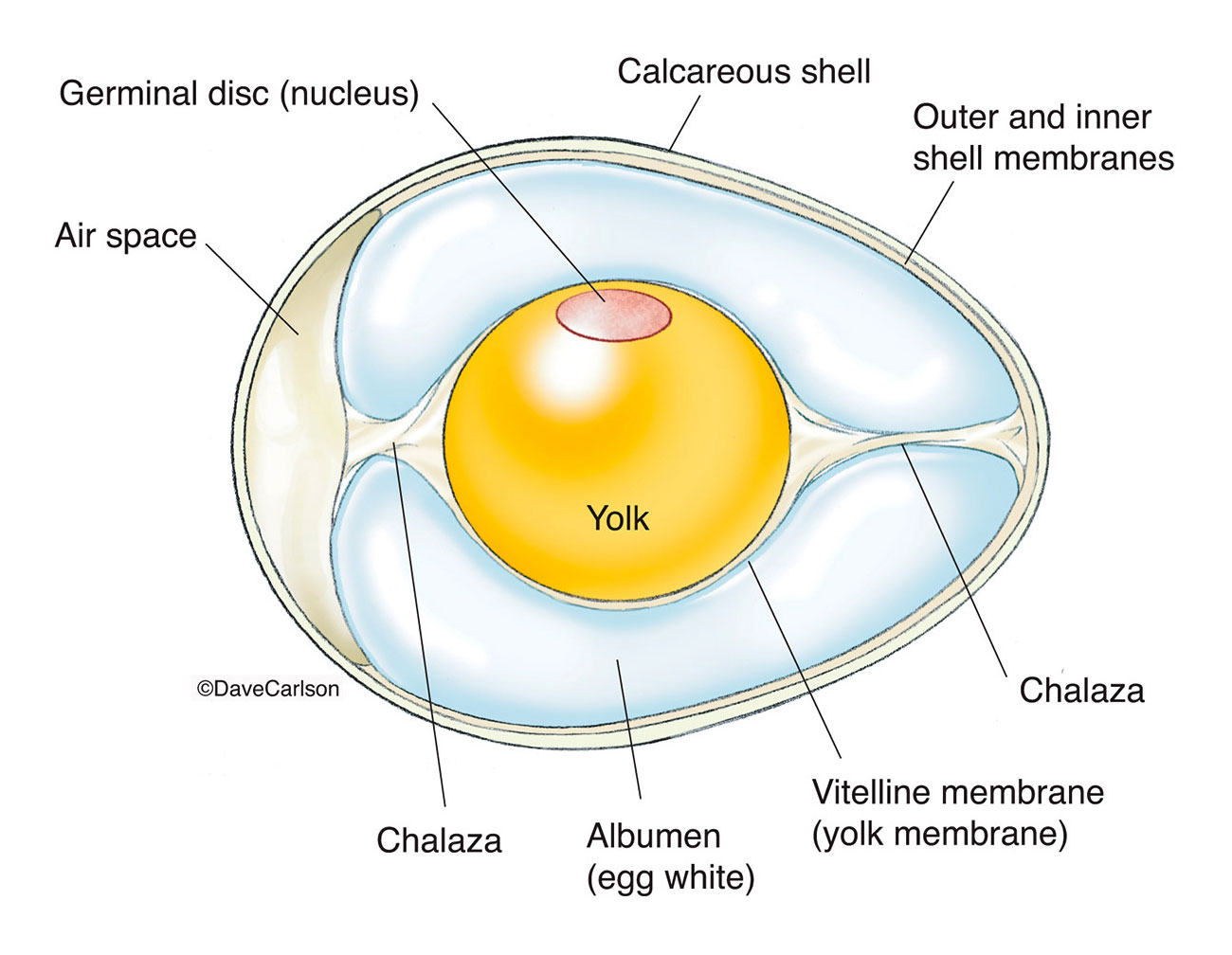
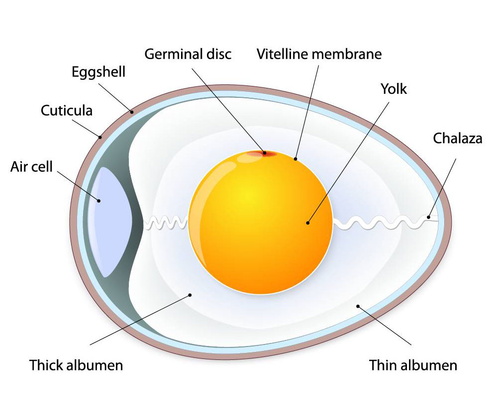
Here is a brief description of the parts of a chicken egg as labeled in a typical diagram:
- Shell: The outermost layer of the egg that provides protection to the developing embryo.
- Shell membranes: Two thin, transparent layers located beneath the shell that help protect the egg from bacteria.
- Air cell: A pocket of air located at the rounded end of the egg that forms as the egg cools after being laid.
- Chalaza: Two twisted, rope-like structures that anchor the yolk to the center of the egg.
- Vitelline membrane: A thin membrane that encloses the yolk.
- Yolk: The yellow, spherical structure that contains the nutrients needed for the developing embryo.
- Germinal disc: A small, circular, slightly raised area on the surface of the yolk that contains the female genetic material.
- Albumen (egg white): The thick, clear, viscous liquid that surrounds the yolk and provides cushioning and protection to the embryo.
The chicken egg is a complex biological structure that provides all of the nutrients and protection needed for the developing embryo to grow and mature into a chick.