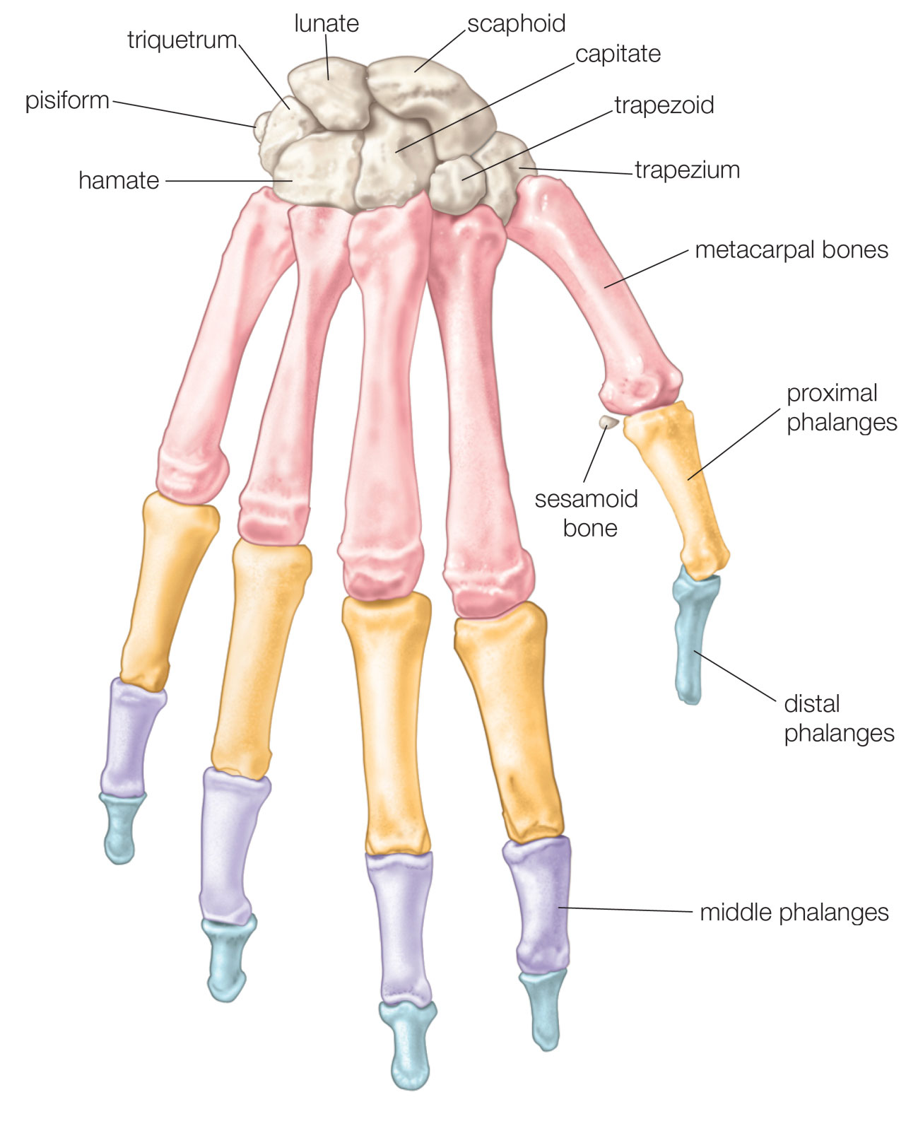
- Distal phalanx: This is the tip of the finger bone that forms the fingertip.
- Middle phalanx: This bone is located between the proximal and distal phalanges.
- Proximal phalanx: This bone is the first bone from the finger tip.
- Metacarpal bone: This is the bone that connects the finger to the wrist.
- Proximal interphalangeal joint (PIP joint): This joint connects the proximal and middle phalanges.
- Distal interphalangeal joint (DIP joint): This joint connects the middle and distal phalanges.
- Carpometacarpal joint (CMC joint): This joint connects the metacarpal bone to the wrist.
- Metacarpophalangeal joint (MCP joint): This joint connects the metacarpal bone to the proximal phalanx.
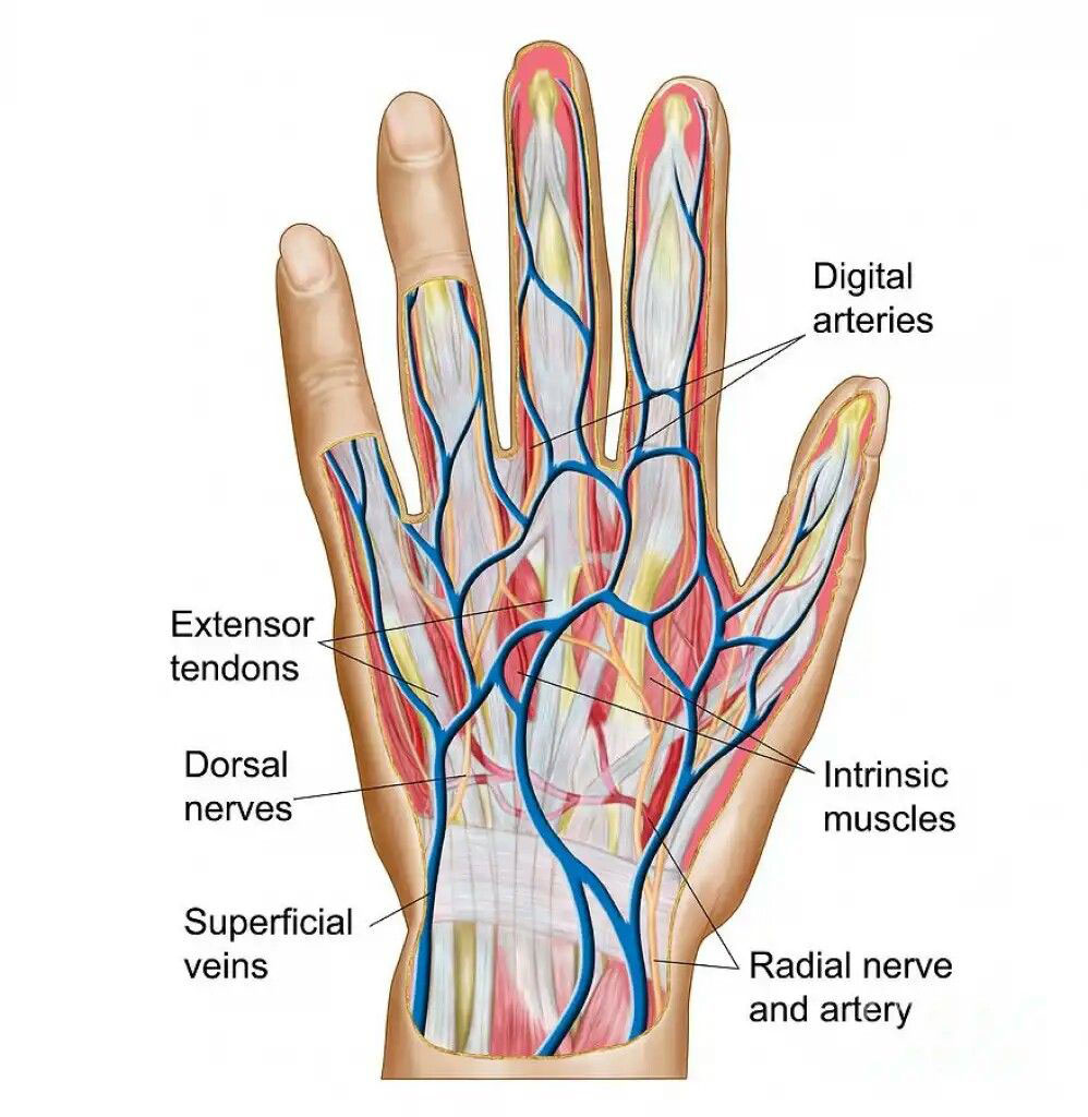
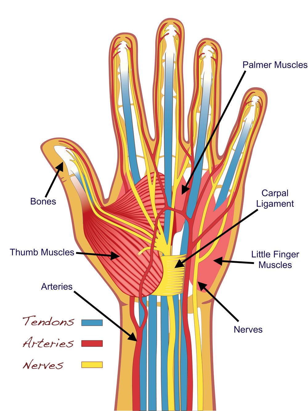
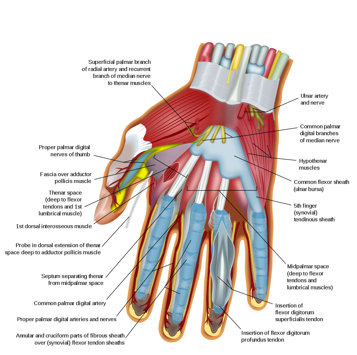
- Flexor tendons: These tendons connect the forearm muscles to the finger bones, allowing the fingers to bend.
- Extensor tendons: These tendons connect the forearm muscles to the finger bones, allowing the fingers to straighten.
- Nail bed: This is the tissue beneath the nail that supports and nourishes the nail.
- Nail plate: This is the hard, protective covering of the fingertip.
- Lunula: This is the half-moon-shaped area at the base of the nail plate.
- Eponychium: This is the fold of skin at the base of the nail plate that covers the matrix, where new nail cells are formed.
- Hyponychium: This is the tissue beneath the free edge of the nail.