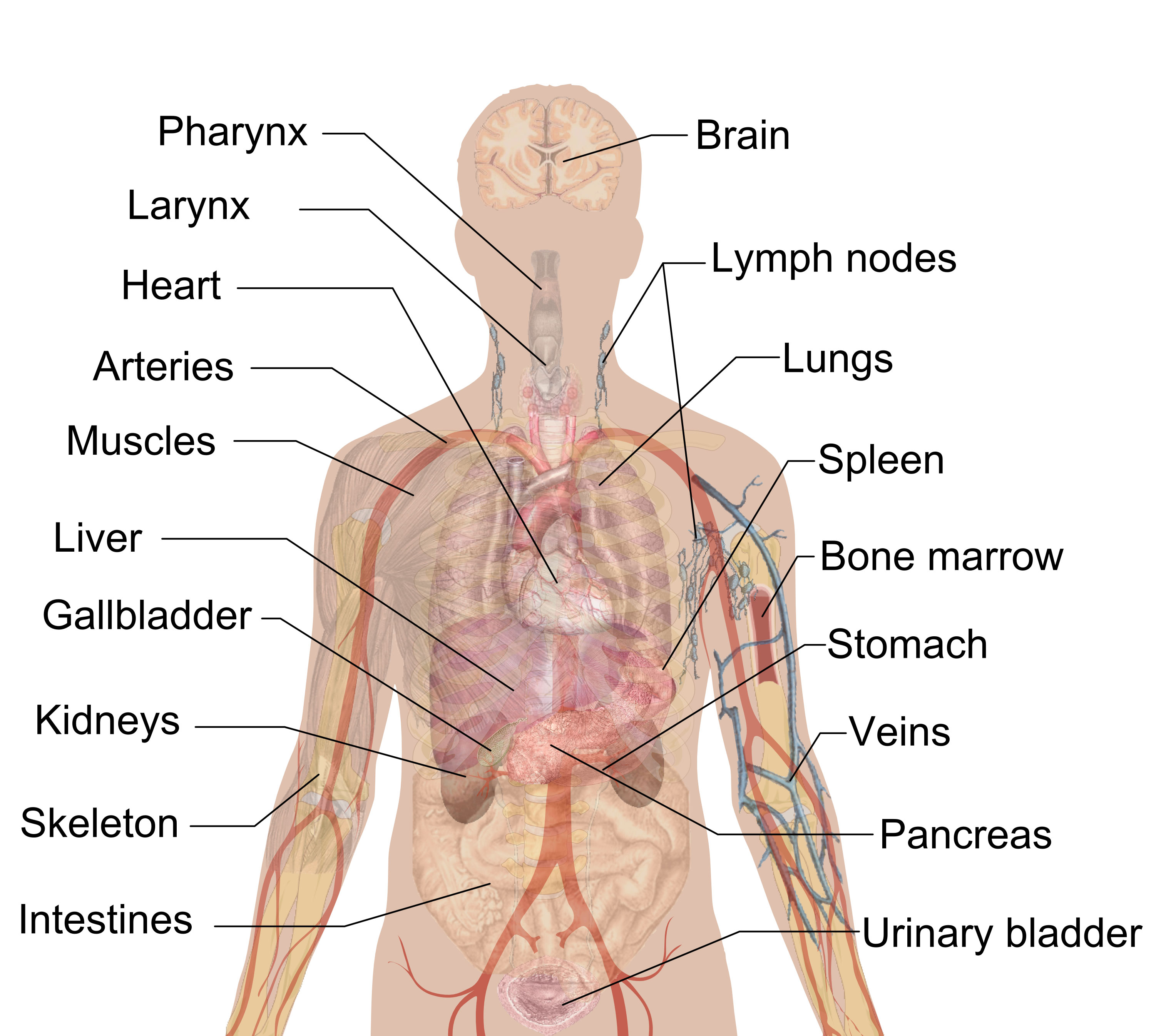
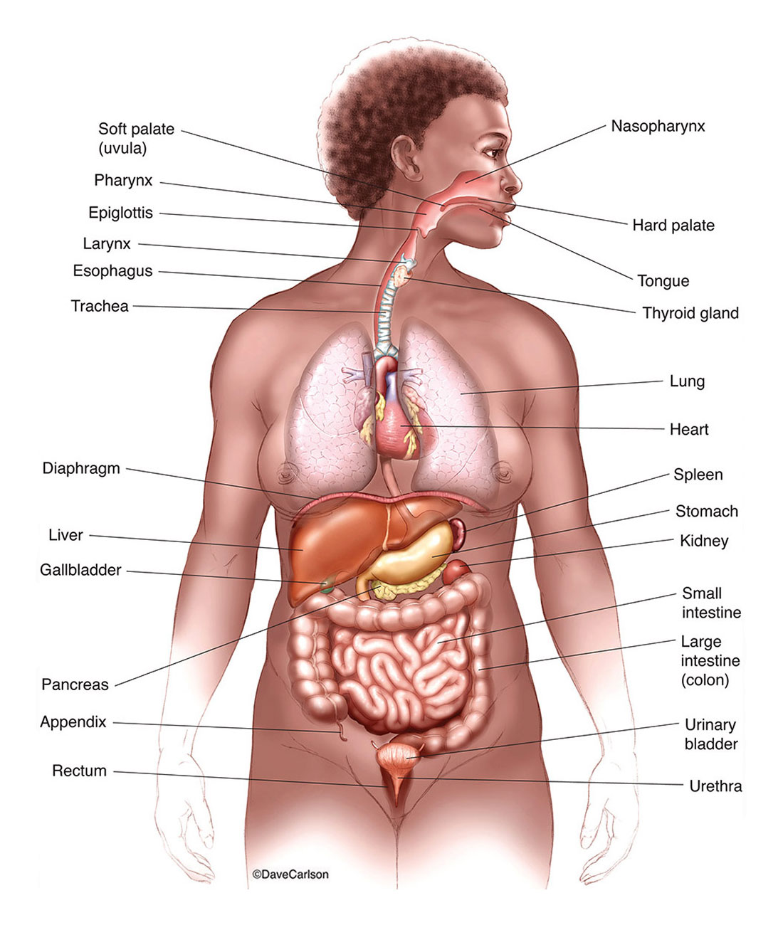
The human internal organs diagram includes the following organs and structures:
- Brain: The control center of the nervous system that is responsible for regulating body functions and controlling behavior.
- Lungs: The respiratory organs that take in oxygen and expel carbon dioxide.
- Heart: The muscular organ that pumps blood throughout the body.
- Liver: The largest glandular organ in the body that produces bile to aid in digestion and detoxifies harmful substances in the blood.
- Stomach: The muscular sac-like organ that mixes and grinds food with digestive enzymes and acid.
- Small intestine: The long, narrow tube where most of the digestion and absorption of nutrients occurs.
- Large intestine: The wider tube that absorbs water and electrolytes from undigested food and forms feces for elimination.
- Kidneys: The pair of bean-shaped organs that filter waste products from the blood and produce urine.
- Bladder: The muscular sac that stores urine until it is eliminated from the body.
- Pancreas: The glandular organ that produces hormones and enzymes to regulate blood sugar levels and aid in digestion.
- Spleen: The organ that helps filter blood and stores white blood cells to fight infection.
- Gallbladder: The small sac-like organ that stores and releases bile to aid in digestion.
- Adrenal glands: The pair of glands that produce hormones that help regulate the body’s response to stress.
- Thyroid gland: The gland that produces hormones that regulate metabolism and growth.
- Reproductive organs: The organs that produce and transport gametes (sperm or eggs) for reproduction, including the ovaries, uterus, fallopian tubes, testes, prostate gland, and penis.