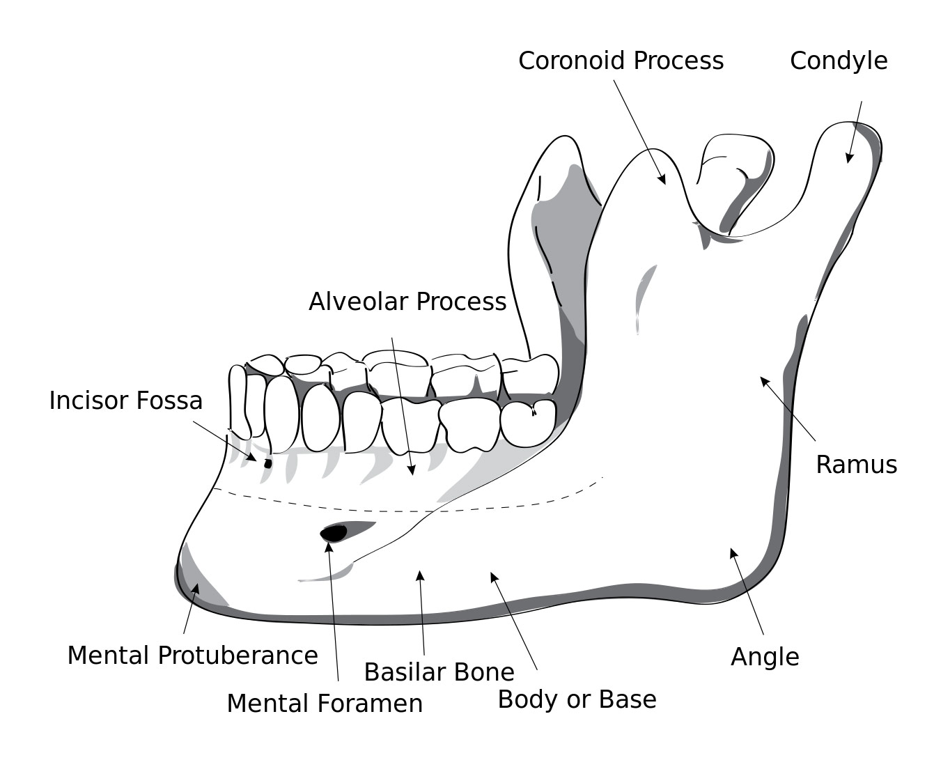
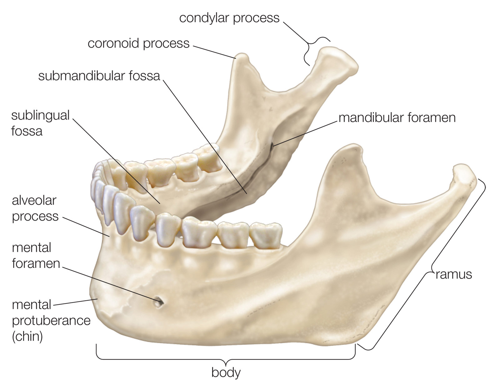
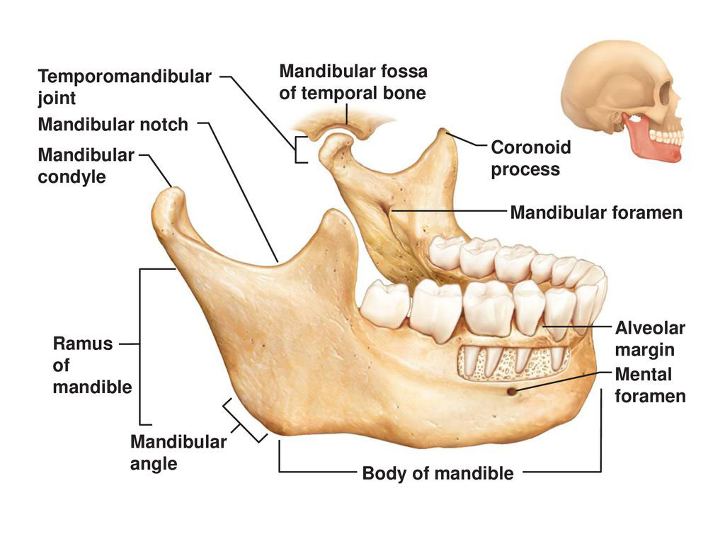
The mandible, commonly known as the jaw bone, is the largest and strongest bone in the human face. A labeled diagram of the mandible bone may include the following parts:
- Body: The horizontal, U-shaped part of the mandible that forms the main body of the bone.
- Ramus: The vertical portion of the mandible that extends upward from the body and forms the side of the jaw.
- Angle: The point where the body and ramus of the mandible meet.
- Condyle: The rounded, knob-like surface at the top of the mandible’s ramus that articulates with the temporal bone to form the temporomandibular joint.
- Coronoid process: A triangular projection at the anterior edge of the mandible’s ramus that serves as an attachment point for the temporalis muscle.
- Alveolar process: The ridge of bone that supports the teeth in the lower jaw.
- Mandibular notch: A groove on the lower border of the mandible that separates the body from the ramus.
- Mental foramen: A small opening on the outer surface of the mandible that allows for the passage of blood vessels and nerves.
The mandible plays a crucial role in the functioning of the human mouth, allowing for biting, chewing, and speaking. The condyle, coronoid process, and alveolar process all serve as attachment points for the muscles that control jaw movement, while the alveolar process supports and anchors the teeth in place. Dysfunction of the mandible can lead to a variety of oral health issues, including misalignment of the teeth, temporomandibular joint disorder (TMD), and jaw fractures.