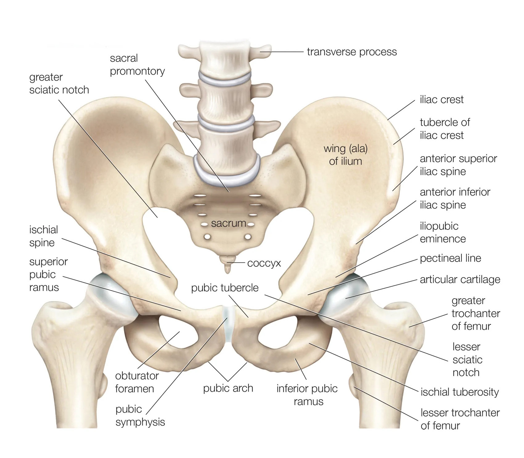
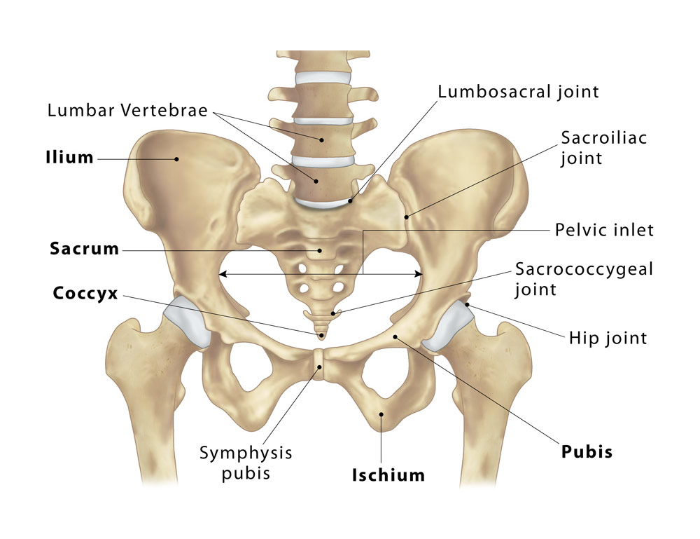
The pelvis is the lower part of the torso, consisting of a group of bones that provide support to the spinal column, protect the internal organs, and aid in movement. It is divided into two halves, the right and left sides, which are joined together by a joint called the pubic symphysis. Here is a brief description of the labeled parts of the pelvis:
- Ilium: The largest bone of the pelvis, which forms the upper part of each hip bone.
- Ischium: The curved bone that forms the lower and back part of the hip bone.
- Pubis: The anterior part of the hip bone that joins with the pubis of the other hip bone at the pubic symphysis.
- Pubic symphysis: The joint where the pubic bones of the two hip bones meet.
- Sacrum: A large, triangular bone located at the base of the spine that forms the back wall of the pelvis.
- Coccyx: A small, triangular bone located at the base of the spine that is often referred to as the tailbone.
- Acetabulum: The cup-shaped socket on the outer surface of the hip bone where the femur (thigh bone) connects to the pelvis.
- Greater sciatic notch: A large notch in the ilium through which the sciatic nerve, the largest nerve in the body, passes.
- Obturator foramen: A large opening in the hip bone that provides passage for nerves and blood vessels.
- Symphysis pubis: The cartilaginous joint that connects the two pubic bones.
These are the main labeled parts of the pelvis. The shape and size of the pelvis can vary between individuals and between genders, and can have implications for childbirth and physical activity.