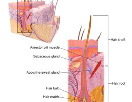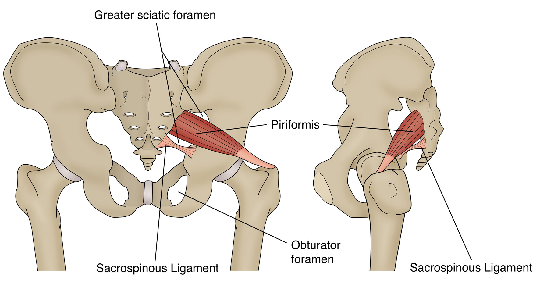
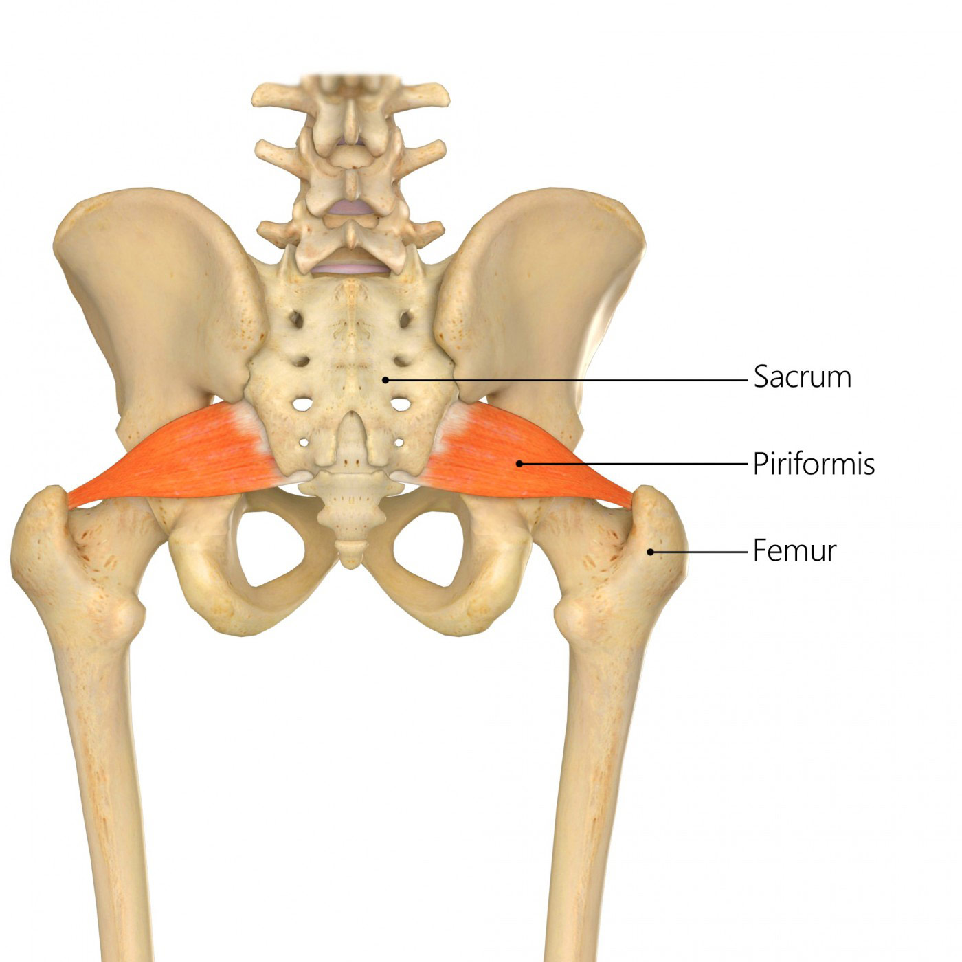
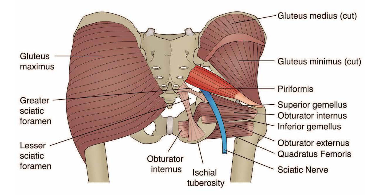
The piriformis is a small, pear-shaped muscle located deep in the buttock region. Here is a brief description of the labeled parts of the piriformis muscle:
- Piriformis muscle: The main muscle of the buttock region, originating from the sacrum (the bone at the base of the spine) and attaching to the femur (the bone of the thigh).
- Sciatic nerve: The largest nerve in the body, which runs from the lower back down to the leg and foot. The sciatic nerve passes under or through the piriformis muscle in some people, which can result in a condition called piriformis syndrome, where the muscle compresses or irritates the nerve and causes pain, numbness or tingling sensations in the buttock and leg.
- Greater trochanter: A bony prominence on the femur, located at the outer aspect of the hip joint and serving as the attachment site for several muscles, including the piriformis.
- Sacrum: The triangular bone at the base of the spine, which forms the posterior part of the pelvis and serves as the origin of the piriformis muscle.
- Femur: The bone of the thigh, which forms the distal (lower) part of the hip joint and serves as the insertion point for the piriformis muscle.
The piriformis muscle plays an important role in hip rotation and stabilization, and is commonly involved in conditions such as piriformis syndrome, hip pain, and sciatica. Understanding the anatomy of the piriformis muscle and its relationship to surrounding structures can be important in the diagnosis and treatment of these conditions.