Skip to content
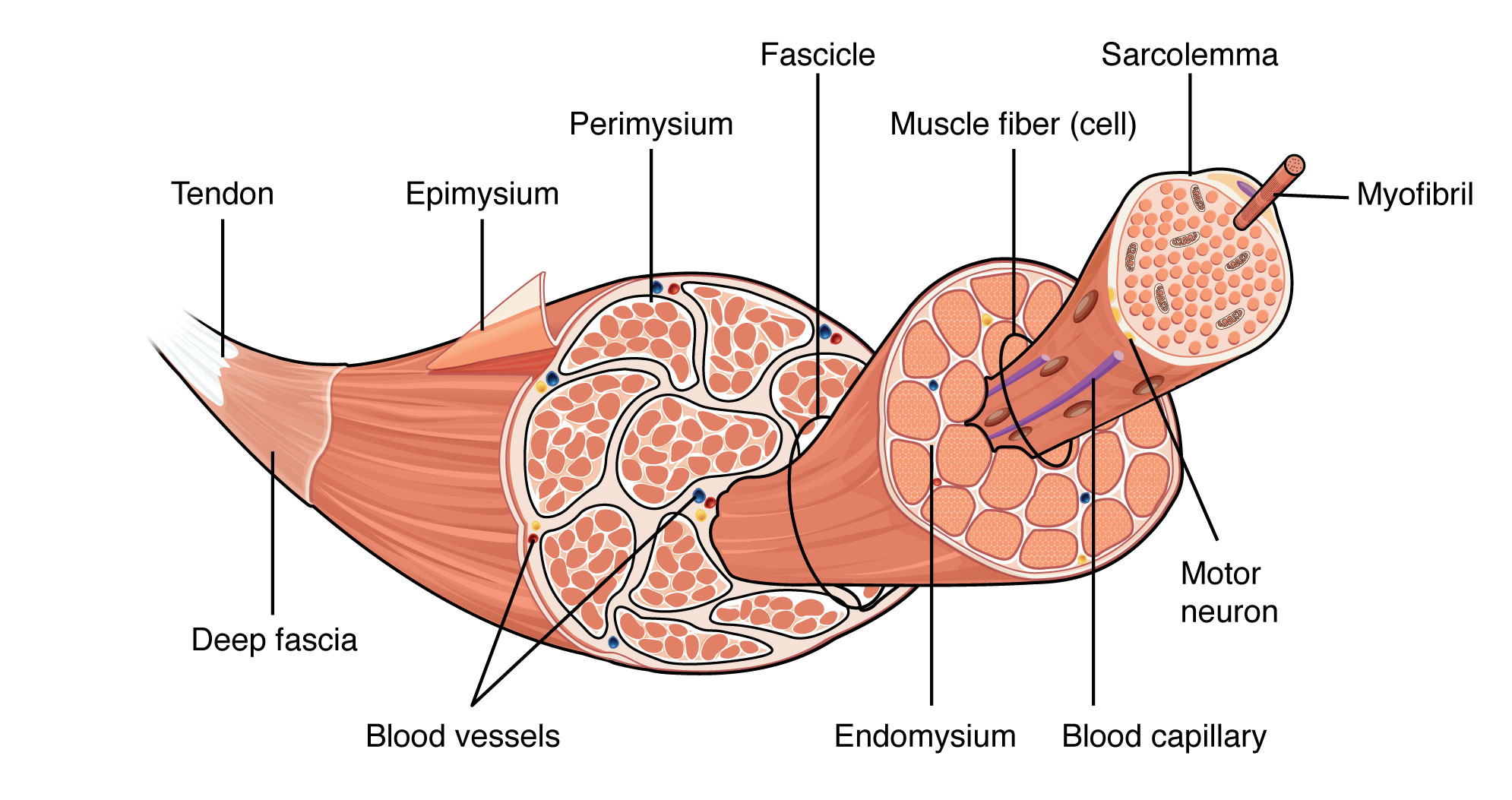
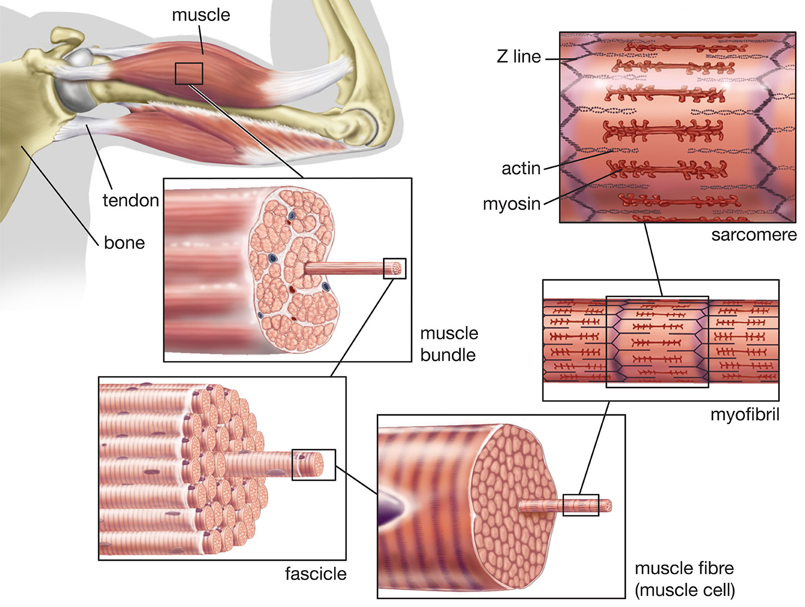
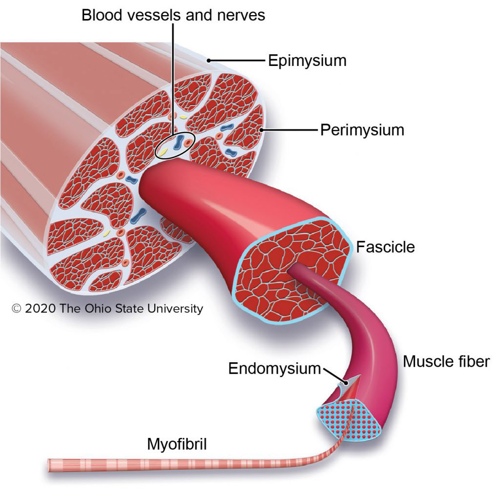
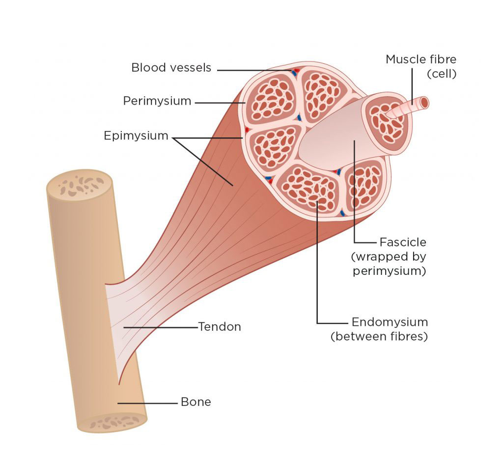
- Epimysium – The epimysium is the outer layer of connective tissue that surrounds the entire muscle.
- Perimysium – The perimysium is a layer of connective tissue that surrounds each bundle of muscle fibers called fascicles.
- Fascicle – A fascicle is a bundle of muscle fibers that are surrounded by perimysium.
- Endomysium – The endomysium is a layer of connective tissue that surrounds each individual muscle fiber.
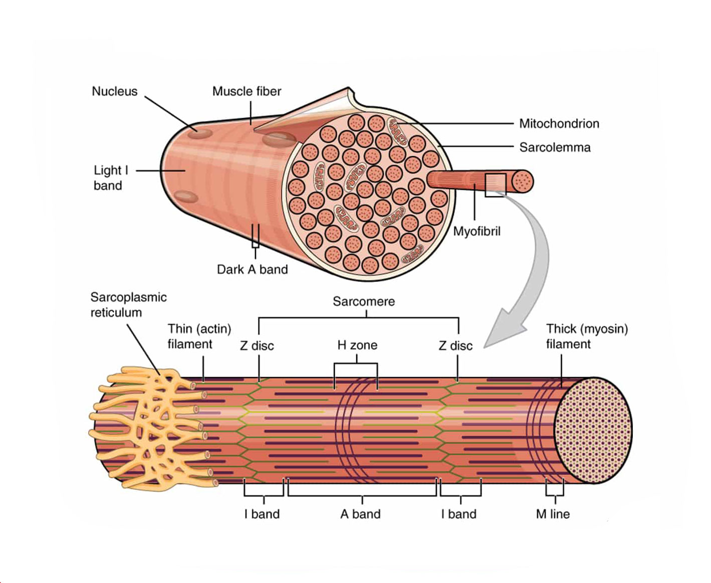
- Muscle Fiber – A muscle fiber is a single muscle cell that can contract and produce force.
- Sarcolemma – The sarcolemma is the plasma membrane that surrounds each muscle fiber.
- Myofibril – A myofibril is a long, cylindrical structure within the muscle fiber that is responsible for producing force.
- Sarcomere – The sarcomere is the functional unit of the myofibril and is composed of actin and myosin filaments.
- Actin Filament – Actin filaments are thin filaments that are responsible for muscle contraction.
- Myosin Filament – Myosin filaments are thick filaments that are responsible for muscle contraction.
- Z-Disc – The Z-disc is a protein structure that anchors the actin filaments in place.
- H-Zone – The H-zone is the central region of the sarcomere where only myosin filaments are present.
- A-Band – The A-band is the region of the sarcomere where both actin and myosin filaments overlap.
- I-Band – The I-band is the region of the sarcomere where only actin filaments are present.
You may also like these posts