Skip to content
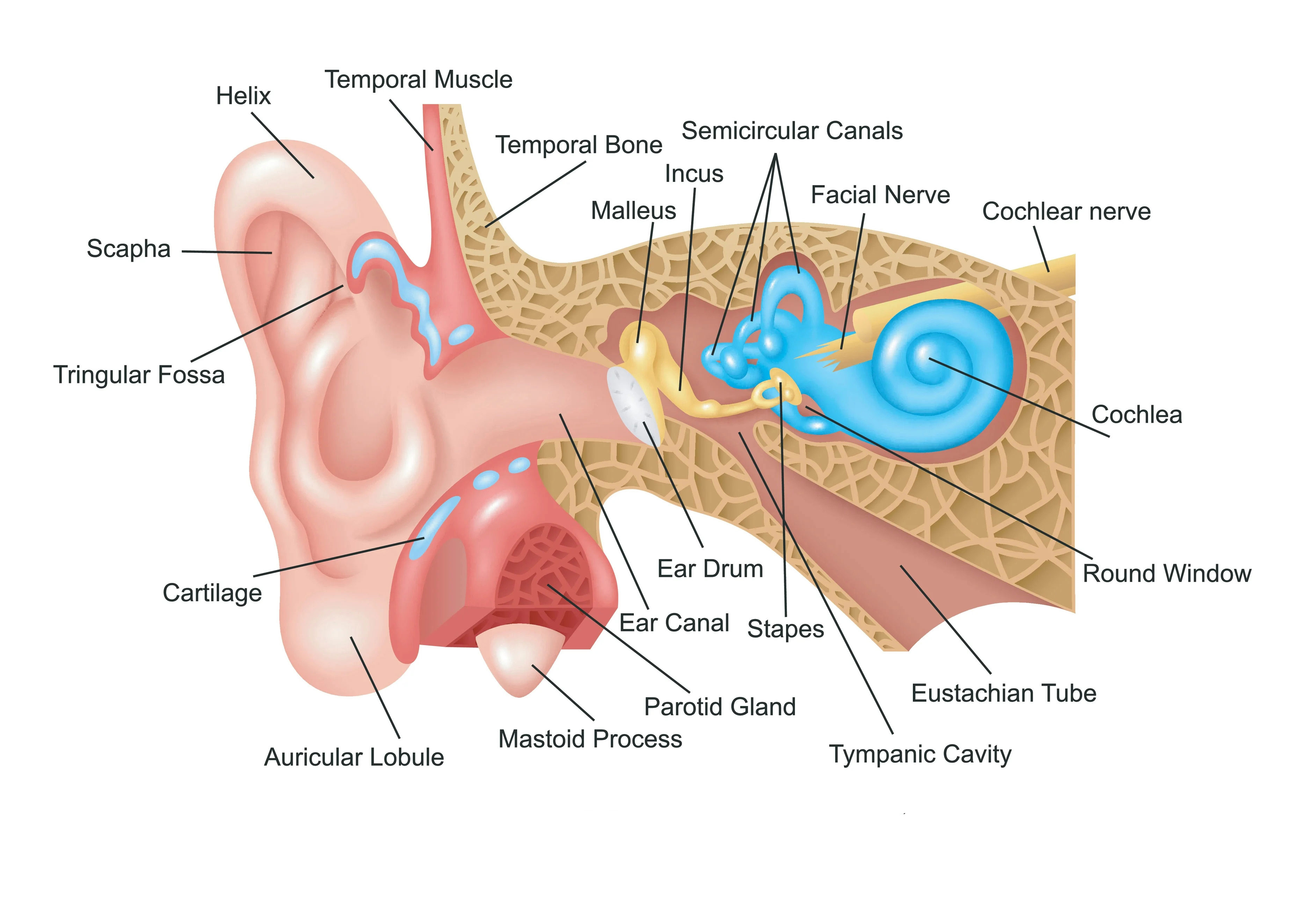
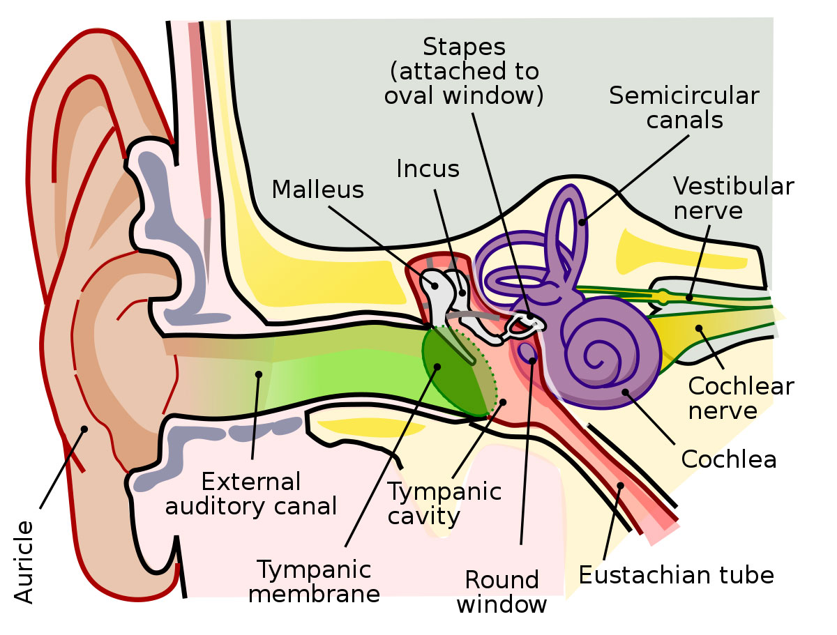
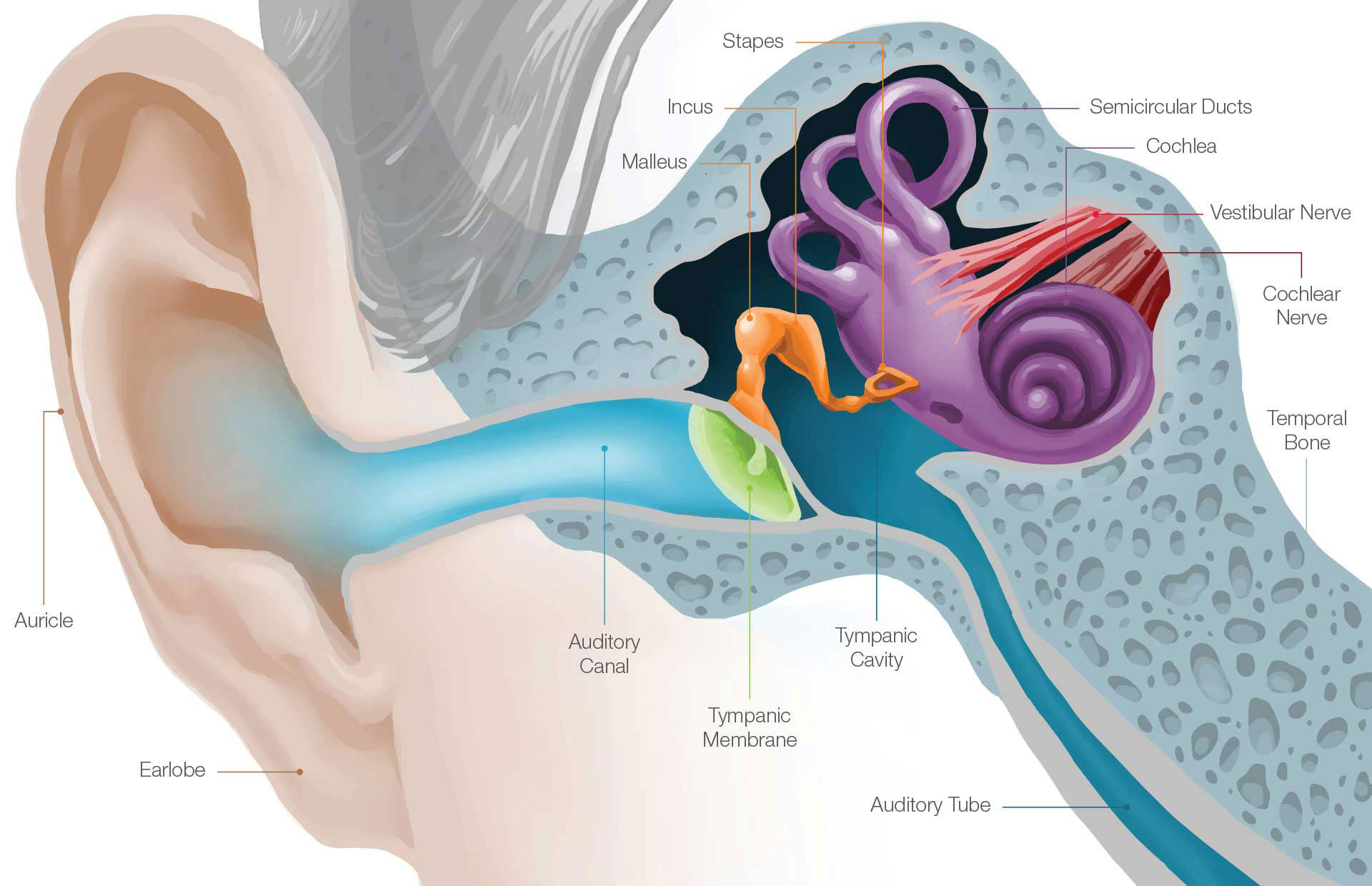
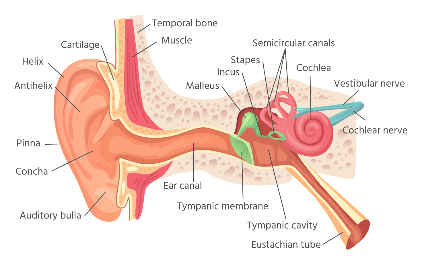
- Pinna: The outermost visible part of the ear which collects sound waves and channels them into the ear canal.
- Ear canal: A narrow tube-like passage that runs from the pinna to the eardrum.
- Eardrum: A thin membrane that separates the ear canal from the middle ear and vibrates in response to sound waves.
- Ossicles: A group of three small bones – malleus, incus, and stapes – located in the middle ear that amplify and transmit the sound waves from the eardrum to the inner ear.
- Eustachian tube: A narrow tube that connects the middle ear to the back of the throat and helps to equalize the air pressure on both sides of the eardrum.
- Cochlea: A spiral-shaped cavity in the inner ear filled with fluid and hair cells that convert sound vibrations into electrical impulses and transmit them to the brain.
- Vestibular apparatus: Consists of three semicircular canals and otolith organs that help to maintain balance and body position.
- Auditory nerve: A bundle of nerve fibers that carries electrical signals from the hair cells in the cochlea to the brain for processing and interpretation of sound.
You may also like these posts