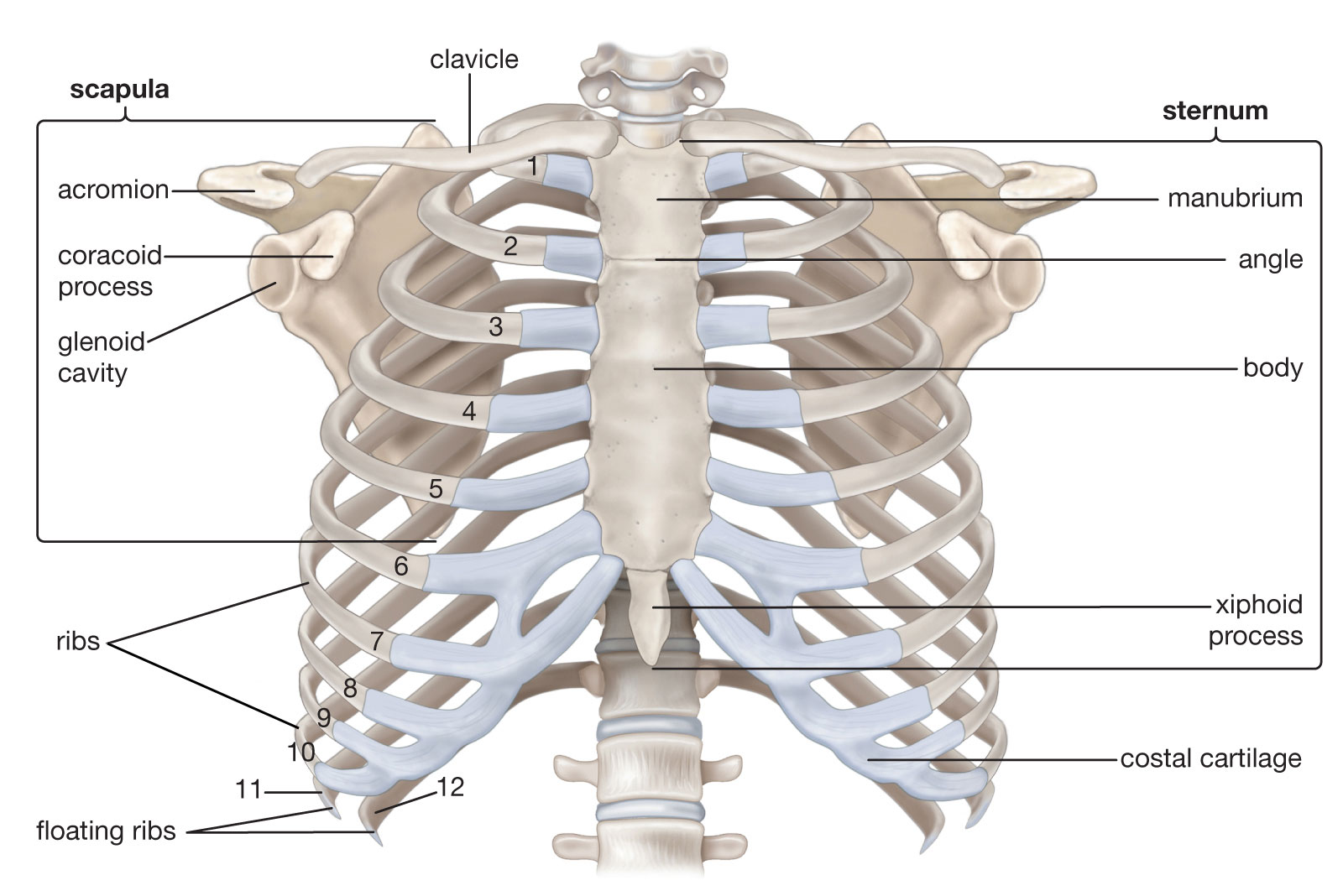
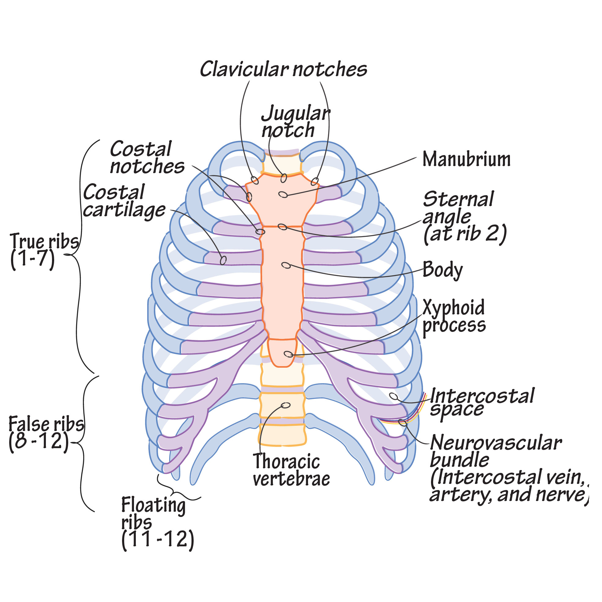
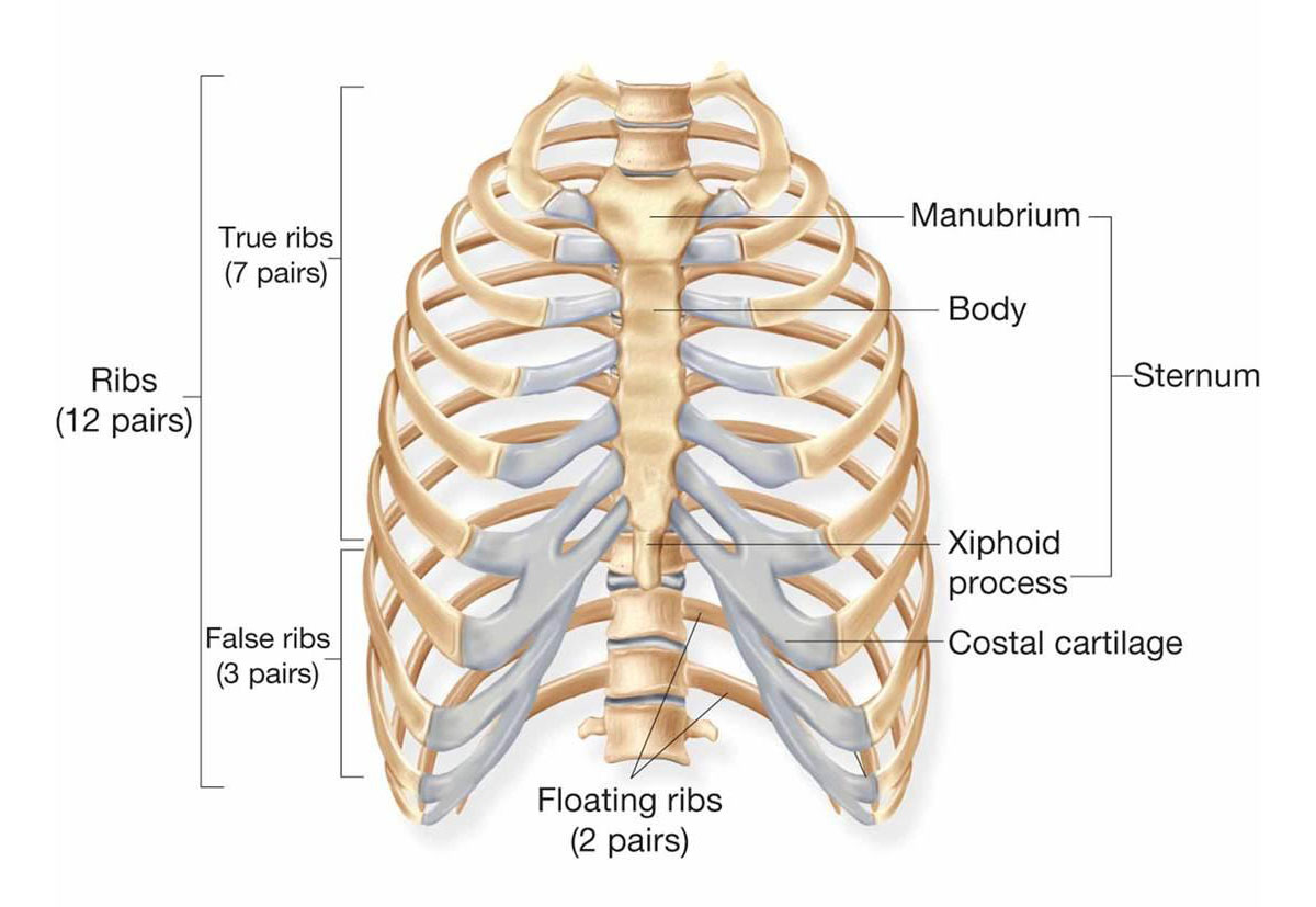
The human thoracic cage, also known as the rib cage, is a bony structure that protects the organs of the chest, including the heart and lungs. Here is a brief description of the parts of the thoracic cage as labeled in a typical diagram:
- Sternum: The breastbone, located at the front center of the thoracic cage, that connects the ribs and provides support for the clavicles (collarbones).
- Clavicle: A long, slender bone located on each side of the sternum that forms part of the shoulder girdle.
- Ribs: Twelve pairs of curved bones that extend from the thoracic vertebrae and wrap around the sides of the chest to meet at the sternum.
- Costal cartilage: The hyaline cartilage that connects the ribs to the sternum.
- Thoracic vertebrae: The 12 vertebrae of the spine that form the posterior part of the thoracic cage.
The thoracic cage is an important part of the human skeleton that provides protection and support for the vital organs of the chest. The movement of the ribs during breathing helps to expand and contract the chest cavity, allowing the lungs to take in and release air.