Skip to content
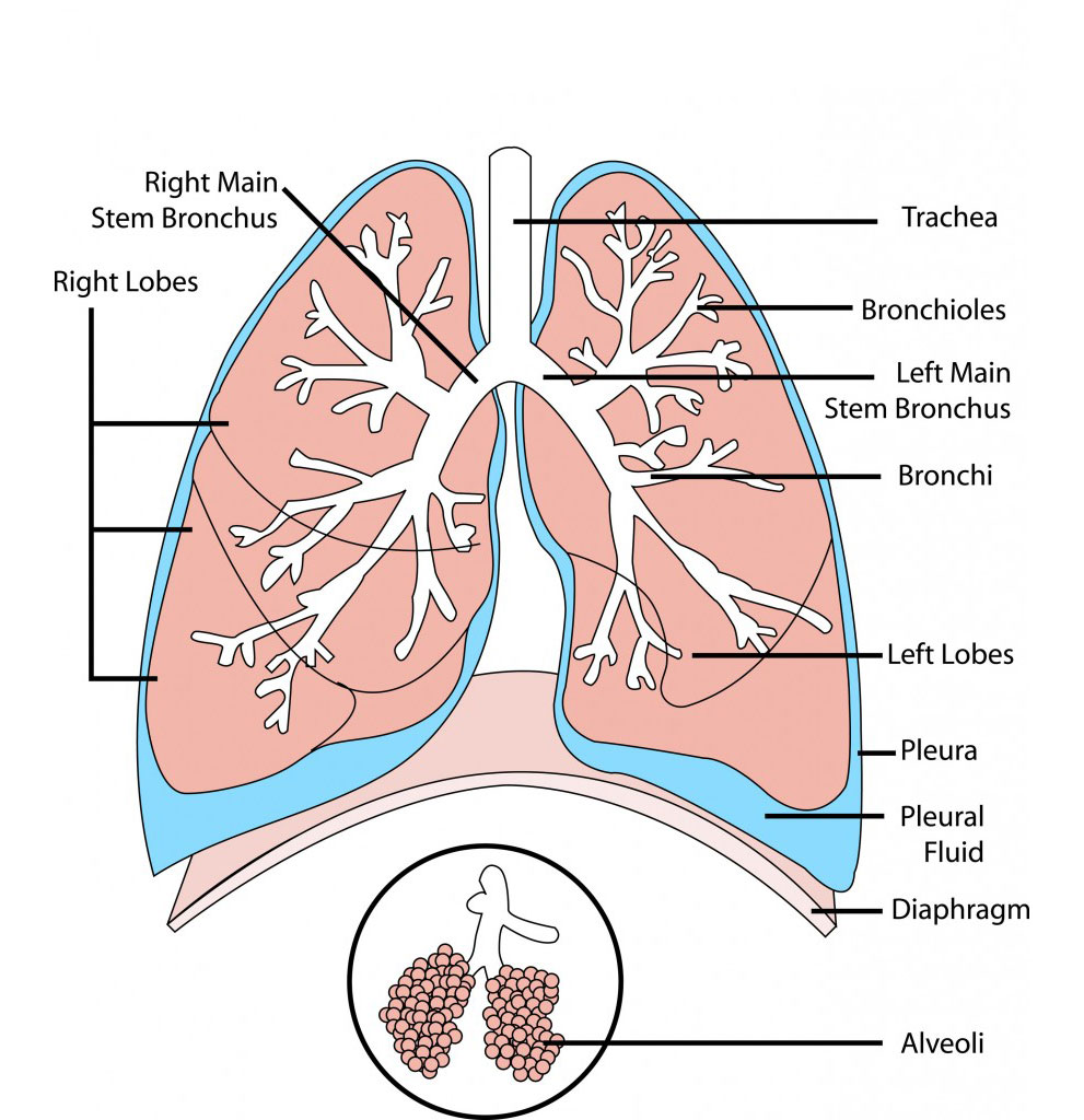
- Trachea: This is the windpipe that connects the lungs to the mouth and nose.
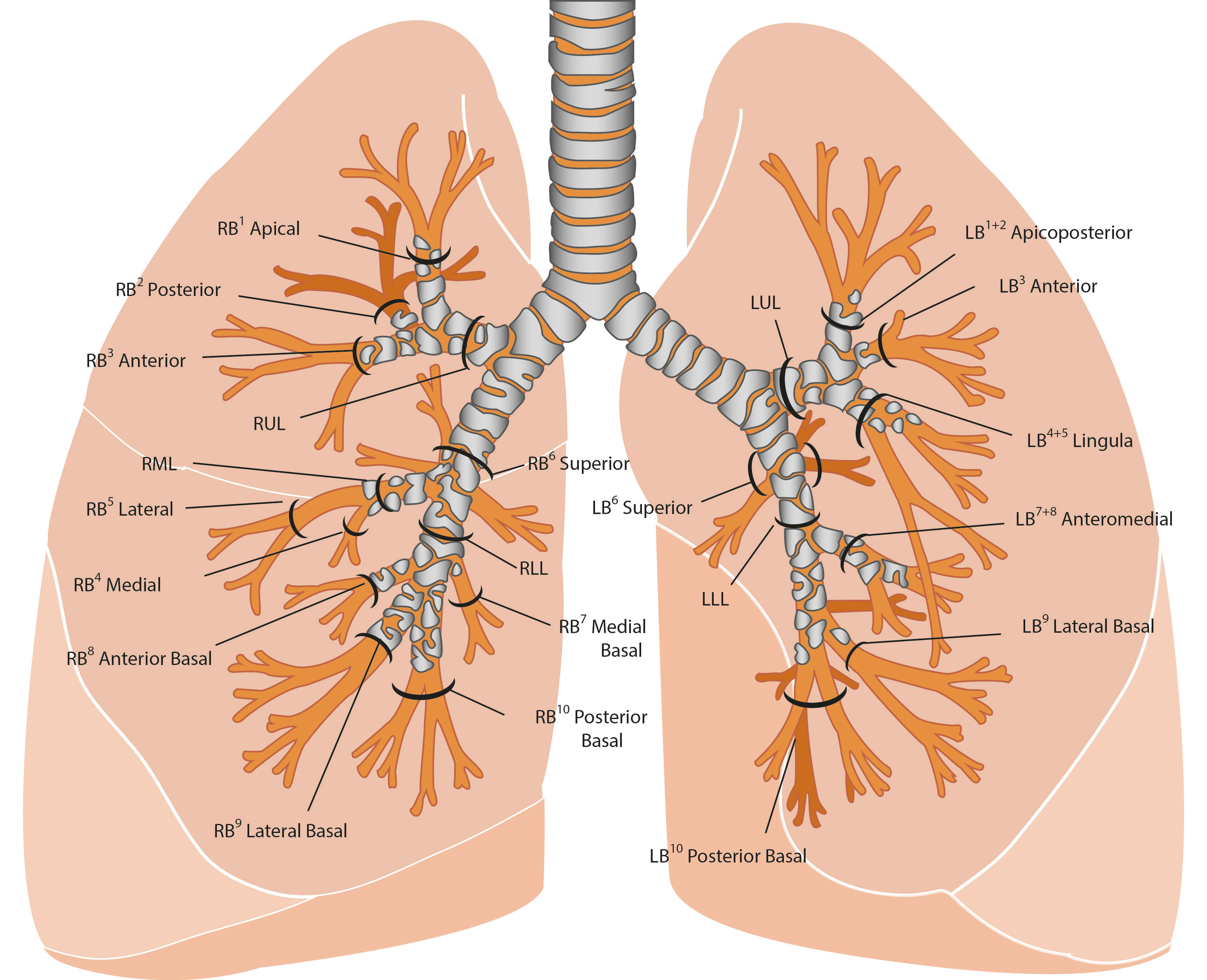
- Bronchi: These are two large airways that branch out from the trachea, one leading to each lung.
- Bronchioles: These are smaller airways that branch out from the bronchi and further divide into tiny air sacs called alveoli.
- Alveoli: These are small sacs where gas exchange occurs, oxygen enters the bloodstream, and carbon dioxide is removed from the body.
- Diaphragm: This is a dome-shaped muscle that separates the chest and abdominal cavity and aids in breathing by contracting and relaxing.
- Pleura: This is a thin layer of tissue that covers the lungs and lines the inside of the chest cavity, helping to reduce friction during breathing.
- Pulmonary veins: These are the veins that bring oxygen-rich blood from the lungs to the heart.
- Pulmonary artery: This is the artery that carries oxygen-poor blood from the heart to the lungs for oxygenation.
- Larynx: This is the voice box that is located at the top of the trachea and contains the vocal cords.
- Pharynx: This is the part of the throat that is behind the mouth and nasal cavity and serves as a passageway for air and food.
- Epiglottis: This is a flap of tissue that covers the trachea during swallowing, preventing food or liquid from entering the lungs.
You may also like these posts